Cool microscopes, nanopatterning and perfusion systems for perturbing and understanding cell signaling dynamics
Many of our projects make use of a range of ultrafast fluorescence microscopes we set up in the lab that can monitor multiple fluorescent reporters at over 500 locations in 96-well plates over several days. These microscopes can be integrated in experiments with automated multiplexed immunostaining for an in-depth analysis of signaling processes in tens of thousands of individual cells after live-cell tracking.
Live-cell imaging to understand cell decision making. Hundreds of thousands of cells are tracked for multiple days and up to 4 fluorescence reporters are measured live in each cell.

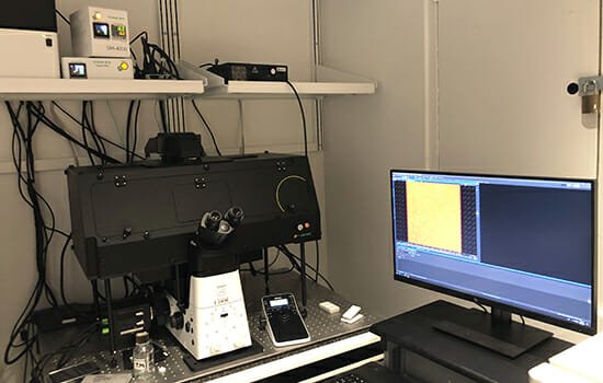
In addition to 5 longterm imaging epifluorescence systems, we have two automated spinning disk microscopes that can image organoids, including one system with super-resolution imaging and one setup for automated local optogenetic manipulations of signaling activites.
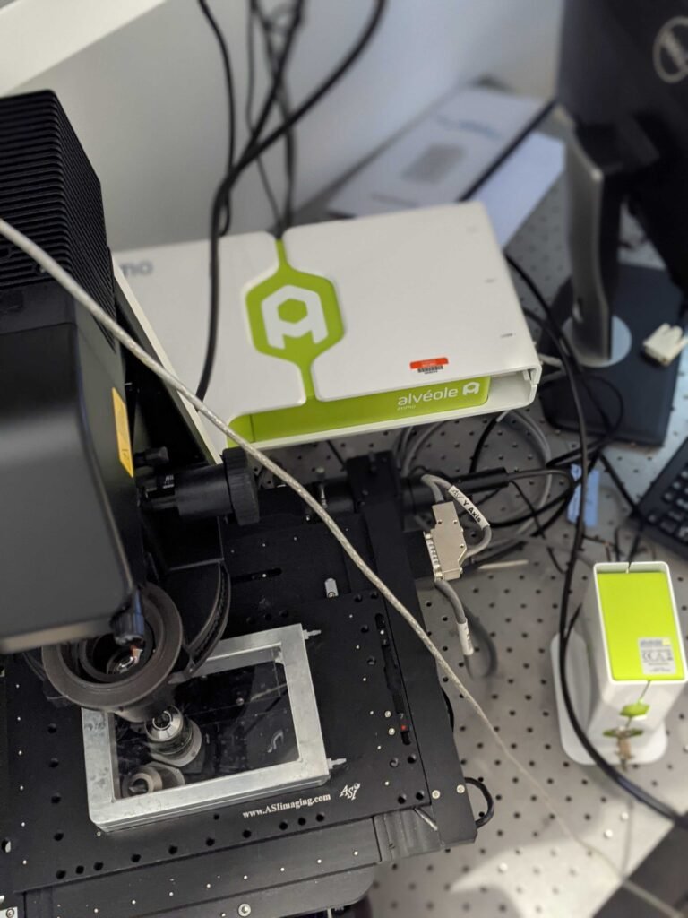
We also have a specialized Primo Alveole microscope system to generate micropatterns in 96-well plates to control where cells grow and move.
Automated cell tracking/quantification